Research Program
1. Enabling technology for musculoskeletal tissue engineering
Tissue engineering tradionally consists of three components: scaffolds, cells and signals. In our lab, we are interested in developing clinically-applicable platform technologies to manipulate scaffolds, cells and signals to create a condition or microenvironment in vitro or in vivo to promote tissue regeneration and self-healing. More specifically, our research aims (1) to integrate microfabrication (bottom-up) with scaffolding (top-down) approaches to re-vascularize engineered cortical and cancellous bones at a large scale, and (2) to achieve temporally and spatially controlled signals that regulate tissue regeneration, leading to a functional tissue regeneration with biomimetic complexity and enhanced functionality. Currently, we are interested in applying the tissue engineering principles to repair and treat musculoskeletal diseases and trauma such as large segmental bone defects, osteonecrosis, rotator cuff injury, and bony birth defects ,along with dental and orthopaedic infections.
One of our major endeavors is to develop bio-inspired biomaterials and medical devices to recapitulate in vivo bony microenvironment. In our lab, we are particularly interested in the concepts of functionally graded biomaterials and various means to realize them by enabling gradual and spatial variation in biomaterial chemistry, structure, property, and signals from nano-, micro- and to macro-level. The goals of our research are to seamlessly integrate different interfacial propertiesand signals and achieve multiple functions. Recently, we have invented a novel bioprinting technology, called Hybprinter, which can seamlessly integrate soft and rigid material components using different printing techniques in a sequential fashion under a single platform. We also developed novel soft and rigid biomaterals that can be used in this hybprinter for acellular and cell-laden medical devices. The technologies we developed in our lab allow us to builid the foundation for vascularized composite tissue constructs, which is potentially a solution for the shortage of organ transplantation and various grafts for disease treatments. Figure 1 shows the schematic of a vascularized composite tissue, the Hybprinter and various representative medical devices and tissue engineering constructs using different modules of the Hybprinter.
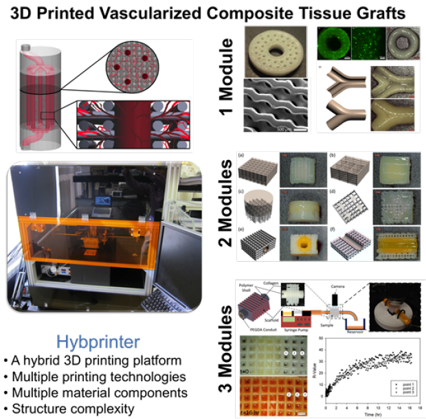
Figure 1 Schematic of vascularized composite tissue constructs, image and features of Hybprinter and representative tissue constructs and medical devices fabricated by Hybprinter.
Rotator cuff injury or tear is the most common injury in shoulder. Re-tear rate for rotator cuff injury even after surgery treatment could be as high as 91%. Some large tear is considered irreparable. Also, the suture anchor technique has been used for more than 100 years for graft fixation to bone, which is though widerly used, but also possesses potential risk for failture due to multiple interfaces. Our lab has recently invented a novel photocrosslinkable polymer, in which we manipulate light exposure to regulate mechanical properties of the polymer to achieve bone-like and tendon-like properties using the same formulation in chemistry. This broad range of mechanical properties in order difference allow us to engineer a functionally graded biomaterial to significantly reduce the interfacial stress concentration to overcome the mechanical mismatch challenge (Figure 2). Furthermore, this unprecedented capability of new biomaterials enables engineering a hybrid suture anchor-tendon graft. This could further reduce the risk of failure by eliminating the suture anchors and graft interfaces by revolutionizing the one century old technique.
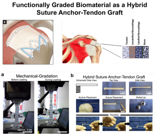
Figure 2 Schematic of repair of large rotator cuff tear using graft, and the bone-tendon interface. (a) A functionally graded biomaterial with bone-like ane tendon-like properties. During loading, the tendon-like side streched, but the bone-like side remained intact. (b) Schematic and rrepresentative images of a hybrid suture anchor-tendon graft and its features.
In addition to hydrogels and polymers, we invented a method to regulate porosity and pore arrangement across a calcium phosphate-based scaffold. Figure 3 shows biodegradable porous bone mineral scaffolds with different pore sizes and spatial arrangements. The porous structure of a calcium phosphate scaffold is critical to facilitate bone regeneration as a template.

Figure 3 Representative digital images of biodegradable porous calcium phosphate scaffolds with different pore sizes and spatial arrangements. A is a scaffold with uniform big pores; B is a scaffold with uniform small pores; C is a scaffold with central small pores and peripheral big pores; D is a scaffold with central big pores and peripheral small pores.
Besides porous structure, the chemistry and signals on the bone mineral scaffold are also very important for bone cells and mesenchymal stem cells. Our recent study was to re-create bony microenvironment with cell-derived extracellular matrix (ECM) and biodegradable β-tricalcium phosphate (β-TCP). More specifically, we investigated whether the ECM produced by bone marrow-derived mesenchymal stem cells (hBMSC) on a β-TCP scaffold can bind bone morphogenetic protein-2 (BMP-2) and control its release in a sustained manner. We further examined the effect of ECM and the BMP-2 released from ECM on cell behavior. The loading and release kinetics of the BMP-2 on the β-TCP/ECM were significantly slower than those on the β-TCP. Furthermore, the BMP-2-loaded β-TCP/ECM stimulated reorganization of the actin cytoskeleton (shown in Figure 4 ), and increased cell expression of alkaline phosphatase and calcium deposition compared to those without BMP-2 loading and the β-TCP with BMP-2 loading.
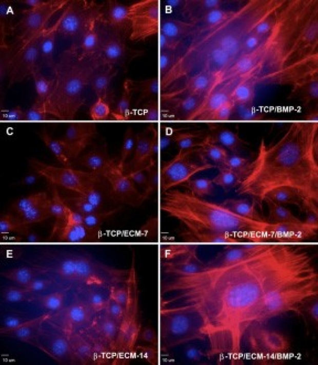
Figure 4 Representative immunofluorescent images for F-actin distribution in hBMSCs taken by a confocal laser scanning microscope after 3 days incubation. (A) Cells in the β-TCP scaffolds group; (B) Cells in the BMP-2-loaded β-TCP scaffolds; (C) Cells in the β-TCP/ECM-7 scaffolds; (D) Cells in the BMP-2-loaded β-TCP/ECM-7 scaffolds; (E) Cells in the β-TCP/ECM-14 scaffolds; and (F) Cells in the BMP-2-loaded β-TCP/ECM-7 scaffolds. (from Biomaterials 2011; 32:6119)
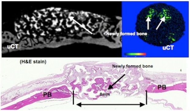
In another study, we evaluated bone formation efficacy aided by biodegradable calcium phosphate scaffolds in the absence of growth factors and stem cells using a rat cranial critical size bone defect (Figure 5 ). Newly-formed bone can be clearly observed by micro CT images and histological stains. We are engineering scaffolds, growth factor delivery, and stem cells for accelerating bone formation.
2. Surface nanotechnology for osseointegrated implant devices
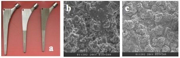

Figure 8 Biological evaluation of plasmas sprayed titanium implants in vitro and in vivo. The left panel shows in vitro osteoblast cells' adhering, migrating and ingrowth on the plasma sprayed titanium porous coating under a scanning electron microscope. The right panel shows osseointegration of new bone-titanium interface under a fluorescent microscope. The plasma sprayed functionally porous graded titanium coated samples were placed into a dog femur. At eight weeks after implantation, the new bone was not only directly in contact with the implant, but also grew into the interconnected pores and formed mechanical interlocking, thereby enhancing the fixation of implants with bone. Yellow indicates new bone; black indicates titanium.
3. Naturally based novel biomaterials for cancer treatment
We are interested in developing naturally based novel biomaterials for improving the efficacy of cancer treatment and reducing side effects. Glioblastoma is the most common malignant tumor of the nervous system in adult humans. The survival rates of patients have not changed in the past 30 years because high grade gliomas mostly recur locally. Therefore, local therapies combined with surgical intervention are an ideal approach to improve the efficacy of treatments. These methods administer drugs directly into the brain, bypass the blood brain barrier (BBB), and deliver the drugs in a concentration-dependent manner. Some clinical studies have demonstrated that local chemotherapy delivered by polymer carrier significantly prolongs the survival time of patients. Currently we are developing a novel naturally based ellagic acid-chitosan composite that is safe and highly efficacious for an improved local chemotherapy.

Figure 9 Fluorescence images of GFP (green fluorescent protein) tagged rat C6 glioma in nude mouse right flanks on the 5th and 21st days after tumor inoculation. The chitosan-ellagic acid composite significantly inhibited the tumor growth in a mouse flank model. (A) tumor-bearing control group; (B) chitosan carrier control group; (C) chitosan-ellagic acid experimental group. The arrows indicate the tumors.